
An Anatomical Study of the Cochlea among Filipinos using High-Resolution Computed Tomography Scans
DOI:
https://doi.org/10.32412/pjohns.v26i1.591Keywords:
cochlea, cochlear turn, high-resolution computed tomography (HRCT), magnetic resonance imaging (MRI)Abstract
Objective: To describe the cochlear anatomy among Filipinos through high resolution computed
tomography (HRCT) imaging.
Methods:
Design: Retrospective Study
Setting: Tertiary Private University Hospital
Patients: Cochlear images retrospectively obtained from computed tomography (CT) scans of subjects who underwent cranial, facial, paranasal sinus and temporal bone computed tomography from October 2009 to July 2010 were reconstructed and analyzed.
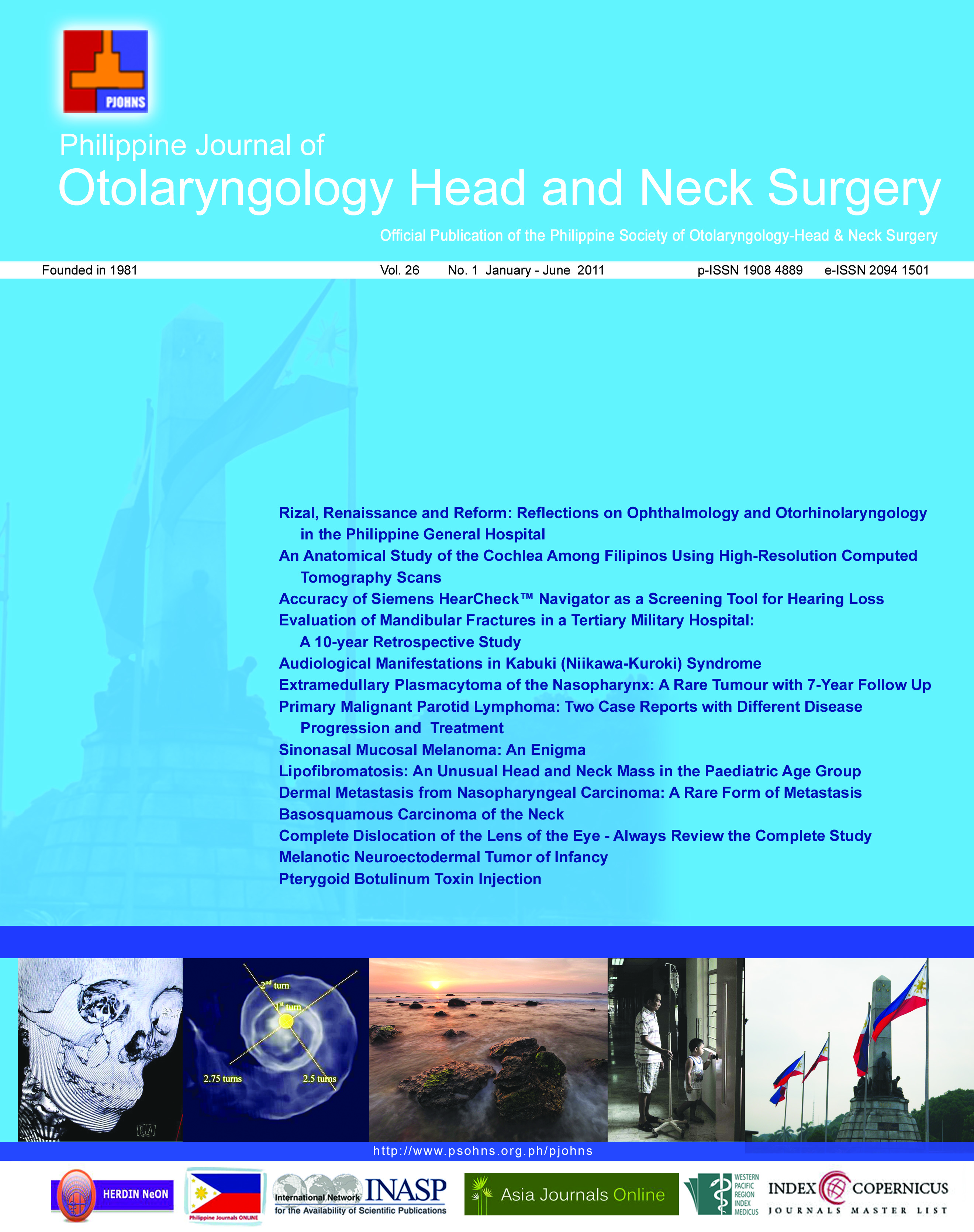
Results: 388 cochlear images were obtained from the scans of 194 subjects (101 males and 93 females, aged 1 to 90 years old, mean = 52 years) and reconstructed for analysis. The mean coiled cochlear height measured 4.36 mm on the right (A.D.) and 4.34 mm on the left (A.S.). Measurement from the oval window to the distal end of the basal turn (equivalent to the horizontal dimension of the cochlea or the mean length of the basal turn) was 7.55 mm A.D. and 7.60 mm A.S. The vertical and horizontal dimensions of right and left cochleas were identical in all subjects (S.D. = 0.35). The right and left cochlear turns were identical in each subject, exhibiting 2 ½ turns in 92.3% of subjects and 2 ¾ turns in 7.7% of subjects.The cochlear dimensions were similar in all subjects, regardless of age. No cochlear ossification or malformation was noted on any CT image.
Conclusion: The 7.55 mm mean length of the cochlear basal turn among Filipinos in this study was 1.24 mm shorter than the average length of the basal turn of 8.81 mm reported elsewhere. Further studies of the cochlear dimensions in specific age groups and its correlation to audiometric status are recommended to determine other significant physiologic correlations.
Keywords: cochlea, cochlear turn, high-resolution computed tomography (HRCT), magnetic resonance imaging (MRI)
Downloads
Published
How to Cite
Issue
Section
License
Copyright transfer (all authors; where the work is not protected by a copyright act e.g. US federal employment at the time of manuscript preparation, and there is no copyright of which ownership can be transferred, a separate statement is hereby submitted by each concerned author). In consideration of the action taken by the Philippine Journal of Otolaryngology Head and Neck Surgery in reviewing and editing this manuscript, I hereby assign, transfer and convey all rights, title and interest in the work, including copyright ownership, to the Philippine Society of Otolaryngology Head and Neck Surgery, Inc. (PSOHNS) in the event that this work is published by the PSOHNS. In making this assignment of ownership, I understand that all accepted manuscripts become the permanent property of the PSOHNS and may not be published elsewhere without written permission from the PSOHNS unless shared under the terms of a Creative Commons Attribution-NonCommercial-NoDerivatives 4.0 International (CC BY-NC-ND 4.0) license.

